Microanalysis using Raman spectroscopy is an invaluable tool for providing high-resolution images of complex molecular samples. Since the Raman effect results from inelastically scattered laser light, this technique requires no labeling or tagging, unlike fluorescence microscopy. Additionally, because of the wavelength independence of the technique, visible laser light can be used providing exceptionally high resolution when compared to other label-free molecular microscopies such as infrared. As a result, Raman microscopy is now commonly used in a wide range of application spaces, including material science, pharmaceuticals, and biomedical.
Unfortunately, despite its popularity in the spectroscopy community, the terms “Raman Microscopy” and “Confocal Raman” are used interchangeably, which often leads to a lot of confusion and miscommunication. While it is true that confocal Raman microscopy is a subset of Raman microscopy the unique optical geometry utilized in a confocal microscope presents both advantages and disadvantages not seen in traditional microscopy. For example, confocal Raman microscopy offers far superior spatial resolution than conventional Raman microscopy, but this comes at the cost of collection efficiency. This white paper sets out to review the fundamental optical design of a confocal Raman microscope, discuss the advantages of this design for collecting Raman spectra, and explore the laser requirements and challenges facilitated by this geometry.
Optical Principals of Confocality
For an optical system to be considered confocal, by definition, it must contain at least two pinholes placed at conjugate locations. In optics, the term conjugate refers to any two locations within the system that have an object / image relationship. It is important to note that this does not mean that these must be located at the actual object and image planes. Most complex optical systems contain several intermediate foci, which may result in multiple conjugates throughout the optical system. Before further diving into the importance of conjugation, it is helpful to take a step back and understand the role of a pinhole in an optical system. Ignoring the wave properties for a second and solely focusing on a geometric (ray) analysis of the pinhole it can be shown that by precisely aligning a pinhole at an intermediate focus in an optical system it will serve to block any off-axis or out of focus rays, while allowing the on-axis to pass through. The smaller the pinhole diameter, the harder it will be to align, but the better the blocking will be. Figure 1 below shows an example of how a properly placed pinhole can block off-axis rays (red) while allowing the on-axis rays (green) to propagate through the system unmolested.
Figure 1: Illustration of a pinholes ability to block off-axis rays when placed at an intermediate focus.
When these pinholes are located at conjugate locations inside of a confocal microscope, they not only block off-axis and out of focus rays, but they significantly reduce the depth of focus of the system allowing for extremely fine z-resolution. This improvement is because the microscope will only pass through rays from the sample plane that is conjugate with both of the pinholes. Allowing for the ability to depth profile transparent samples and / or produce structured opaque samples. In a confocal Raman microscope, the first pinhole is placed in the laser path. This pinhole serves the dual purpose of establishing the first conjugate point and providing a spatial filter to clean up the beam, ensuring a diffraction limited spot at the sample. The second pinhole is then placed in front of the spectrometer, which if properly aligned, will prevent any unwanted light collected, scattered, or emitted by the sample from entering the detector. Figure 2 below shows a simplified diagram where the excitation laser (green) is focused through the first pinhole, then recollimated and directed to the objective lens with a dichroic mirror. When the laser light is focused into the sample, there will be Raman scattering not only from the exact location where the beam is focused (red) but also slightly above and below (black). While all of these rays may fall within the depth of focus of the microscope objective, the pinhole in front of the spectrometer limits the rays which are capable of propagating into the spectrometer so only the ones which are from the sample plane conjugate with the first pinhole in the laser path.

 SHIPS TODAY
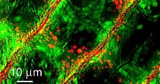
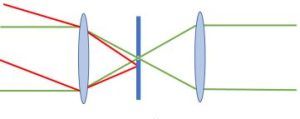 Figure 1: Illustration of a pinholes ability to block off-axis rays when placed at an intermediate focus.
When these pinholes are located at conjugate locations inside of a confocal microscope, they not only block off-axis and out of focus rays, but they significantly reduce the depth of focus of the system allowing for extremely fine z-resolution. This improvement is because the microscope will only pass through rays from the sample plane that is conjugate with both of the pinholes. Allowing for the ability to depth profile transparent samples and / or produce structured opaque samples. In a confocal Raman microscope, the first pinhole is placed in the laser path. This pinhole serves the dual purpose of establishing the first conjugate point and providing a spatial filter to clean up the beam, ensuring a diffraction limited spot at the sample. The second pinhole is then placed in front of the spectrometer, which if properly aligned, will prevent any unwanted light collected, scattered, or emitted by the sample from entering the detector. Figure 2 below shows a simplified diagram where the excitation laser (green) is focused through the first pinhole, then recollimated and directed to the objective lens with a dichroic mirror. When the laser light is focused into the sample, there will be Raman scattering not only from the exact location where the beam is focused (red) but also slightly above and below (black). While all of these rays may fall within the depth of focus of the microscope objective, the pinhole in front of the spectrometer limits the rays which are capable of propagating into the spectrometer so only the ones which are from the sample plane conjugate with the first pinhole in the laser path.
Figure 1: Illustration of a pinholes ability to block off-axis rays when placed at an intermediate focus.
When these pinholes are located at conjugate locations inside of a confocal microscope, they not only block off-axis and out of focus rays, but they significantly reduce the depth of focus of the system allowing for extremely fine z-resolution. This improvement is because the microscope will only pass through rays from the sample plane that is conjugate with both of the pinholes. Allowing for the ability to depth profile transparent samples and / or produce structured opaque samples. In a confocal Raman microscope, the first pinhole is placed in the laser path. This pinhole serves the dual purpose of establishing the first conjugate point and providing a spatial filter to clean up the beam, ensuring a diffraction limited spot at the sample. The second pinhole is then placed in front of the spectrometer, which if properly aligned, will prevent any unwanted light collected, scattered, or emitted by the sample from entering the detector. Figure 2 below shows a simplified diagram where the excitation laser (green) is focused through the first pinhole, then recollimated and directed to the objective lens with a dichroic mirror. When the laser light is focused into the sample, there will be Raman scattering not only from the exact location where the beam is focused (red) but also slightly above and below (black). While all of these rays may fall within the depth of focus of the microscope objective, the pinhole in front of the spectrometer limits the rays which are capable of propagating into the spectrometer so only the ones which are from the sample plane conjugate with the first pinhole in the laser path.
